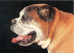
Certain breeds of dogs and cats are prone to difficult, obstructive breathing because of the shape of their head, muzzle and throat. The most common dogs affected are the “brachycephalic” breeds. Brachycephalic means “short-headed.” Common examples of brachycephalic dog breeds include the English bulldog, French bulldog, Pug, Pekingese, and Boston terrier. These dogs have been bred to have relatively short muzzles and noses and, because of this, the throat and breathing passages in these dogs are frequently undersized or flattened (Figure 1). Persian cats also have a brachycephalic conformation.
The term Brachycephalic Syndrome refers to the combination of elongated soft palate, stenotic nares, and everted laryngeal saccules, all of which are commonly seen in these breeds.
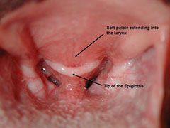
Elongated soft palate (Figure 2) is a condition where the soft palate is too long so that the tip of it protrudes into the airway and interferes with movement of air into the lungs.
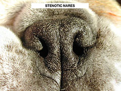
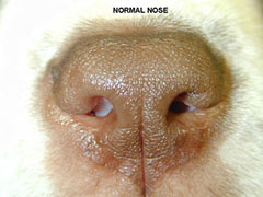
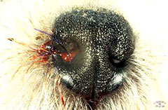
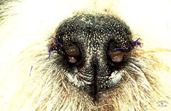
Stenotic Nares (Figure 3a) are malformed nostrils that are narrow or collapse inward during inhalation, making it difficult for the dog to breathe through its nose.
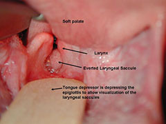
Everted Laryngeal Saccules (Figure 4) is a condition in which tissue within the airway, just in front of the vocal cords, is pulled into the trachea (windpipe) and partially obstructs airflow.
Some dogs with brachycephalic syndrome may also have a narrow trachea (windpipe), collapse of the larynx (the cartilages that open and close the upper airway), or paralysis of the laryngeal cartilages.
Dogs with elongated soft palates generally have a history of noisy breathing, especially upon inspiration (breathing inward). Some dogs will retch or gag, especially while swallowing. Exercise intolerance, cyanosis (blue tongue and gums from lack of oxygen), and occasional collapse are common, especially following over-activity, excitement, or excessive heat or humidity. Obesity will aggravate the problems. Many dogs with elongated soft palates prefer to sleep on their backs. This is probably because this position allows the soft palate tissue to fall away from the larynx. The signs associated with stenotic nares and everted laryngeal saccules are similar.
Stenotic nares can be easily diagnosed on physical examination (Figure 3). Definitive diagnosis of both elongated soft palate and everted laryngeal saccules can only be made with the dog under anesthesia. Generally, brachycephalic breeds have a thick tongue that makes visualization of the larynx in an awake animal very difficult. Attempts to restrain the patient and retract the tongue sufficiently to allow visualization of the larynx are generally unsuccessful. Under anesthesia, elongated soft palates extend past the tip of the epiglottis (the entrance to the airway). In severe cases the soft palate will extend directly into the laryngeal opening. The tip of the soft palate and the edges of the larynx are often inflamed (swollen and red). In chronic cases, the cartilages of the larynx become inflexible and begin to collapse, further narrowing the airway. Everted laryngeal saccules look like blue-gray soft tissue masses protruding into the airway just in front of the vocal folds (Figure 4). Your primary care veterinarian may also recommend chest x-rays to evaluate your pet’s lower airways and lungs.
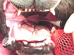
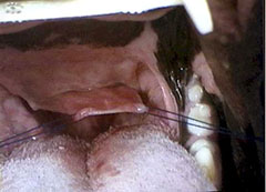
Soft palate abnormalities should be treated if they cause distress to your pet, become more severe with time, or cause life-threatening obstruction. If your pet shows gagging, coughing, exercise intolerance, or difficulty breathing, resection of the excess soft palate may be necessary. Soft palate resection (staphylectomy) is performed using a scalpel blade, scissors, or CO2 laser. (Figure 5) The palate is stretched (Figure 6) and the excess tissue is removed with blade or scissors.
If the laryngeal saccules are everted, they may be removed at the same time as the soft palate resection, or they may be left in and allowed to return to a more normal position. Correction of stenotic nares, if present, helps improve breathing and is done at the same time (Figures 7 and 8).
Pets must be monitored very closely immediately after surgery. Significant inflammation or bleeding can obstruct the airway, making breathing difficult or impossible. Occasionally a tube must be placed and maintained through an incision in the neck into the trachea (temporary tracheostomy) until the swelling in the throat subsides enough that the pet can breathe normally.
Pets are usually observed in the hospital for at least 24 hours. Post-operative coughing and gagging are common. In chronic cases in which the laryngeal cartilages have become inflexible, removal of the elongated soft palate and laryngeal saccules may not provide enough relief. The creation of a new permanent opening into the trachea in the neck area (called a permanent tracheostomy) may be the only solution, although there are complications associated with this procedure as well.
The prognosis is good for young animals. They generally will breathe much easier and with significantly reduced respiratory distress. Their activity level can markedly improve. Older animals may have a less favorable prognosis, especially if the process of laryngeal collapse has already started. If the laryngeal collapse is advanced, the prognosis is poor.


