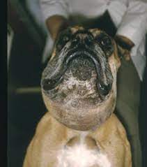
A salivary mucocele, or sialocele, is a collection of saliva that has leaked from a damaged salivary gland or salivary duct, and has accumulated in the tissues. This is often noted as a fluctuant, painless swelling of the neck or within the oral cavity. While often inaccurately called a salivary cyst, mucoceles are lined by inflammatory tissue (called granulation tissue) which is secondary to the inflammation caused by the free saliva in the tissues. A cyst is lined by epithelial (glandular) tissue which is itself responsible for the production of the fluid. Salivary mucoceles may be classified as follows:
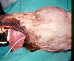
- Cervical Mucocele: This is the most common type of mucocele. It is a collection of saliva in the upper neck region, under the jaw, or in the intermandiublar region (between the jaws). (Figures 1 and 3)
- Sublingual Mucocele (also called a ranula): Another frequent location for the formation of a mucocele is on the floor of the mouth alongside the tongue. This is frequently seen in association with a cervical mucocele. (Figure 2)
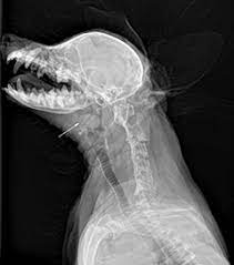
- Pharyngeal Mucocele: This type of mucocele is much less common. It is essential a variation of the cervical mucocele, but the fluid accumulation is almost entirely within the throat (pharynx). (Figure 4)
- Zygomatic Mucocele: This is a very rare type of mucocele where the saliva is originating from the small zygomatic salivary glands which are located just below the eye.
The cause of salivary mucoceles is rarely identified, although trauma such as from choke collars, bite wounds, or chewing on foreign materials is generally considered to be the most likely initiating event. As the saliva leaks from the torn salivary gland or duct, it accumulates in the tissue and initiates an intense inflammatory response. A connective tissue capsule gradually forms around the saliva to prevent it from migrating further.
This condition is almost exclusively seen in dogs and very rarely in cats. All breeds are susceptible but there seems to be an increased incidence in Poodles, German Shepherds, Dachshunds, and Australian Silky Terriers. There is no age predisposition and this condition may occur at any time.
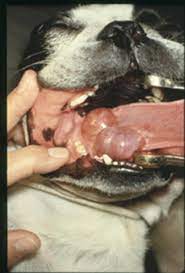
Generally the development of a cervical mucocele (Figure 1) is seen as a gradually enlarging soft, painless, fluctuant mass in the upper cervical (neck) or intermandibular region. In most dogs and cats there are no problems associated with the development of the mass. If the mucocele is a sublingual mucocele (ranula) (Figure 2), your pet may have some difficulty eating and may develop bleeding from trauma to the mucocele as he or she chews. A pharyngeal mucocele is generally totally undetectable until the oral cavity is examined with sedation. Pets with pharyngeal mucoceles may experience respiratory distress because the mass developing in the throat is beginning to obstruct the airway. This is a potentially very serious problem, and treatment must be instituted rapidly because these pets may die from acute respiratory distress. Difficulty swallowing may be another sign that a pharyngeal mucocele is present.
Generally, the diagnosis of a salivary mucocele is fairly straightforward. Palpation of the salivary glands in normally easily accomplished, and, with the exception of the pharyngeal mucocele, the mucoceles are easily identified as a soft, fluctuant swelling that is non-painful. Tumors and abscesses may appear similar but are generally either firm or painful.
Occasionally cervical mucoceles migrate to the ventral midline over time, making it difficult to determine whether the problem involves the left or right sided glands (Figure 1). Examining the pets with sedation on their back (Figure 3) often allows the mucocele to migrate to the affected side.
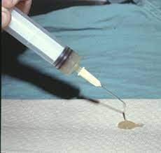
Laboratory abnormalities are generally not helpful in the diagnosis of salivary mucocele. If uncertainty exists about whether the mass is a mucocele or an abscess, a sterile aspiration of the fluid can be performed. Aspiration of a clear, yellowish or blood-tinged thick ropy fluid with a low cell count is consistent with saliva (Figure 5). An elevation of the white blood cell count in the fluid may indicate an infection in the salivary gland (sialadenitis) or an abscess. Sometimes, special laboratory testing (staining) can help confirm the type of fluid if there is doubt.
Radiographs are rarely needed to diagnose salivary mucoceles; however, if neoplasia (cancer) is suspected x-rays of the thorax are indicated to look for metastasis.
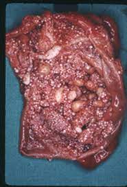
The only satisfactory treatment for a salivary mucocele is removal of the salivary gland or glands that are involved with the mucocele. (Figure 6)
Continued aspiration of a mucocele will not permanently eliminate the problem. It will occasionally resolve the problem for weeks to several months, but most will recur. Aspiration also risks introducing bacteria into the mucocele, which can potentially cause an infection that will significantly increase the difficulty of successful surgical treatment.
Surgical removal of the mandibular and sublingual glands on the side of the mucocele is the normal surgical treatment. The glands are removed together because the duct of the mandibular gland travels through the sublingual gland and removal of one gland would unavoidably traumatize the other. The mandibular gland is closely associated with the large veins that join to form the jugular vein. Removal of the salivary glands requires careful dissection to the area of several critically important nerves.
Sublingual mucoceles (ranulas) (Figure 2) may be treated with marsupialization (in addition to removal of the mandibular and sublingual glands) to facilitate drainage into the oral cavity. Marsupialization is performed by excising an elliptical portion of sublingual mucosa overlying the mucocele and suturing the rim of oral mucosa to connective tissue.
Frequently a drain is placed in the area of the mucocele to allow fluid to escape from the area until it has a chance to heal.
If a drain was left in the surgical site, your pet will experience several days of drainage. If the wound is bandaged, it will be necessary to change the bandage frequently. If the wound is not bandaged, it is helpful to apply warm compresses with a damp towel. This will help clean the skin in the area of the surgery and will help encourage drainage of fluid from the area.
Prognosis is excellent for a normal life after drainage of a mucocele and adequate removal of the affected salivary glands. Dogs do not suffer from a dry mouth following removal of the mandibular and sublingual glands, even if performed on both sides.
Postoperative complications are uncommon if the procedure is performed by an adequately trained surgeon. Occasionally a fluid pocket (seroma) may develop in the area where the mucocele was. This can either be drained or may be allowed to resolve by itself. Infections are possible but uncommon. If inadequate glandular tissue is removed, it is possible that the mucocele will recur.


