Coxofemoral luxation is the dislocation of the hip joint resulting in displacement of head of the femur from the acetabular socket. Dislocation produces disruption of the joint capsule and other supportive structures of the hip, including ligaments and often bone. Hip luxation is commonly caused by trauma, but joint degeneration or hip dysplasia increases the risk.
Traumatic hip luxation is painful, impacting weight bearing and leg function. Leg carriage is dramatically altered by hip luxation. The leg is often tucked, outwardly rotated, and shortened or inwardly rotated and deviated dependent on the direction the hip is dislocated. In the majority of cases (90%), the femoral head is displaced forward and above the acetabulum (the hip socket). Partial dislocation of the hip joint (subluxation) can occur and is typically associated with joint degeneration as with hip dysplasia. Subluxation is commonly bilateral whereas bilateral hip luxation is rare.
A full general examination and orthopedic evaluation should be performed due to the possibility of concurrent trauma. The force required to produce hip luxation can also damage the urinary system, lungs, heart, or other body organs. Additional diagnostics are needed to assess the patient for other injuries or organ conditions which may affect anesthesia. Additional blood work is indicated to fully assess organ function, identify the extent of damage, identify pre-existing medical problems, and for anesthetic planning.
- Bloodwork
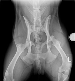
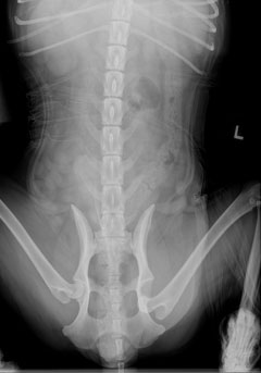
- Radiographs (x-rays) of the hips are performed to assess direction of dislocation and other associated damage to the joint such as fracturing of articular surfaces (Figures 1 and 2). Heavy sedation or general anesthesia may be needed.
- Additional radiographs (chest, spine, abdomen or other limbs to assess for concurrent trauma)
1) Non-surgical reduction of the hip (closed reduction): In a closed reduction the hip is replaced under short anesthesia and often supported in this position with a sling. Slings cannot always be applied due to limb trauma or leg conformation that doesn’t tolerate sling placement. If used, slings must be monitored closely for rub sores or changes in position. If reduction is maintained for several weeks to support tissues to heal, then surgery is avoided with good results. Closed reduction is successful approximately 50% of the time.
2) Surgical reduction: Treatment consists of surgically replacing the hip (open reduction) and restoring the supporting structures. Additional supporting implants are usually placed to help mechanically support the hip during the healing phase. There are a large variety of techniques that may be chosen based on your ACVS board-certified veterinary surgeon’s experience, including toggle rod fixation, surgical anchors, reconstruction of the joint capsule, and trochanteric transposition. Success is high with most of these techniques.
3) Femoral Head Ostectomy (FHO): Restoration of the hip is often not feasible due to injury or poor hip conformation. The femoral head and neck ostectomy removes the femoral head and neck producing a “false joint.” Function is very good with this technique although there is a mild loss in function. With proper patient selection, good functional results are expected with this option. Postoperative physical therapy is of great benefit in maintaining good function after FHO. The risk of recurrent luxation or implant complication is eliminated with this technique.
4) Total hip replacement: In this scenario the joint is replaced with synthetic materials. Several hip replacement alternatives are available, which consist of replacing the femoral head and the acetabulum (ball and socket) with synthetic implants.
Healing of the supporting structures of the hip requires several weeks and your pet’s activity will need to be restricted for at least six to eight weeks. Longer periods of restriction may be needed for certain procedures. Following healing of regional supporting hip structures, re-strengthening of the limb musculature will require an additional period of controlled progressive activity. These guidelines will need to be individually modified by your primary care veterinarian through your pet’s recovery.
Aftercare will also be customized for both rate of recovery and for the specific repair performed (recovery after femoral head ostectomy may be accelerated). Coexisting injury, patient physique, and other ailments will also influence recovery recommendations. Controlled activity with exercises is often started by two weeks following surgery.
Most complications are not life threatening, but can be nonetheless frustrating. These include re-luxation of the hip; implant complications; and bandage and sling complications. Complications delay recovery and can limit ultimate functional outcome. Be mindful that even the best patients are not fully cooperative with exercise restrictions and uncomfortable slings. Failure can occur with a simple unlucky fall or slip during the recovery process. Post-treatment activity can cycle implants causing them to migrate and break. Slings used for immobilizing the limb can migrate producing severe injury to the leg and must be monitored carefully.
Prognosis is excellent for eventual return of good-excellent limb function depending on the severity of injury or underlying degeneration. Additional treatment may be required for failed closed hip reduction requiring further surgical repair. Historically approximately 50% of closed hip reductions and 10-20% of open reduction require further surgical repair. The requirement of revision is not necessarily a failure of a surgical technique as patient condition, associated injuries, hip architecture, patient compliance and aftercare all play a role. Persistence and dedication eventually produce good functional outcome in the majority of patients.
Premier Sponsors