The liver performs an incredible number of functions to maintain health of animals, including filtering out toxins, storing sugar, and making proteins. Most of the blood that is carried to the liver for these processes arrives via the portal vein, which drains the intestines, stomach, pancreas, and spleen. Within the liver, the portal vein branches into smaller and smaller vessels so that the blood can percolate throughout the tissues to each liver cell. When these microscopic vessels are abnormal on liver biopsy, the condition is called “hepatic microvascular dysplasia (HMD or MVD)” or “portal atresia”. When the microscopic vessels within the liver are underdeveloped or absent, the liver becomes small (“atrophied”) and the animal can no longer process toxins or make proteins necessary for growth and normal function.
Hepatic microvascular dysplasia (HMD) or portal atresia is a histologic diagnosis, meaning it only describes the biopsy findings. In fact, there are many conditions that can cause these findings, including congenital portosystemic shunts; however, when the diagnosis is made without evidence of a congenital shunt, then the dogs are often given the diagnosis of HMD as a specific disease.
Dogs with HMD can present with signs and blood works with abnormalities similar to dogs with congenital portosystemic shunts; however, many dogs have no clinical signs at all. Often affected dogs are 3 to 4 years old before they have clinical signs. Some affected dogs are smaller than normal, with poor muscle development. They may seem less intelligent or quieter because of the toxins that depress their brains. They may have a loss of appetite or occasional bouts of vomiting and diarrhea. Some dogs may have a greater risk of infections or develop bladders stones. Severely affected dogs may be wobbly or act drunk or blind and can even seizure. Rarely, dogs will develop fluid filled bellies from liver failure.
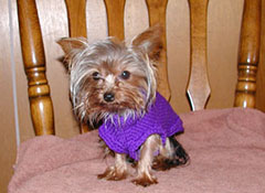
Yorkshire terriers and Cairn terriers are most commonly affected, but the condition is also seen in many other small breeds, including Maltese, dachshund, miniature poodles, Shih tzu, Lhasa apso, cocker spaniel, and West Highland white terriers.
In some dogs, basic biochemical tests are normal. Severely affected dogs may have low blood protein, albumin, glucose, and urea nitrogen levels because their livers are not making enough of these chemicals. Some dogs have increased liver enzymes. Urine is evaluated for evidence of infection and crystals; rarely, dogs with HMD will develop ammonium biurate crystals in the urine that look like spiky balls or starfish.
Bile acids are measured after an overnight fast (“preprandial” or fasting) and then 2 hours after eating (“postprandial”). In dogs with HMD, one or both sets of bile acids are increased. Bile acids can increase with any liver disease, so high bile acids are not specific to congenital portosystemic shunts or HMD.
A definitive diagnosis of HMD is made by proving that the dogs do not have any shunts but that they do have abnormal vessels on their liver biopsies. Dogs with HMD have normal portal blood flow on nuclear scintigraphies (nuclear scans of liver blood flow), portograms (x-ray studies of liver blood flow), and CT angiograms (CAT scans of liver blood flow), but they have abnormal microscopic portal blood vessels on liver biopsy. The liver biopsy is usually taken surgically through a belly incision or with a laparoscope so that enough liver tissue can be obtained to evaluate the blood vessels. Needle biopsy using ultrasound may not provide enough tissue to make the diagnosis.
HMD must be differentiated from congenital portosystemic shunts; unfortunately some dogs can have both diseases, and this cannot be determined before surgery (Figure 1). If your dog undergoes surgical closure of a congenital portosystemic shunt and its bile acids remain high 3–6 months after surgery, it is quite possible he/she also had congenital HMD. This is why liver biopsy is recommended at the time of shunt attenuating surgery. Dogs with HMD are usually older than dogs with shunts when they are diagnosed (2 to 5 years instead of less than one year), and often their blood work changes are less severe than dogs with shunts. They may even have normal fasting bile acids, but usually their postprandial bile acids are increased.
There are no surgical treatments for HMD. Dogs with the condition are managed medically, and treatment is based on the severity of the condition. In some dogs no treatment is needed. The mainstay of medical management is to reduce the amount of protein in the diet. Specific veterinary diets such as Hill’s L/d have been formulated for dogs with liver disease. The protein is highly digestible (often milk based or soy) and is only mildly protein restricted. Diets for dogs with HMD should contain about 15–20% protein (roughly 2 g/kg per day of protein), 15-30% fat, and 30-50% highly digestible carbohydrates on a dry matter basis. They should also be high in zinc and Vitamin E and low in manganese. Most dogs with HMD do well on diet change alone.
Changing the type of bacteria that live in the intestines can also decrease toxin production and absorption. This can be accomplished by giving lactulose syrup or yogurt. Your veterinarian may prescribe antibiotics for a short time as well.
Nutriceuticals — compounds that are not considered “drugs” — can also improve liver function. Milk thistle (“Silymarin”) can help improve liver function and regeneration. Because the government does not regulate over-the-counter compounds, purchase of specially formulated veterinary supplements is recommended. Two veterinary companies that sell milk thistle include Nutramax (“Marin”) and RxVitamins (“Hepatosupport”). Veterinarians may also prescribe Denosyl (SAM-e) to improve liver function.
Prognosis is good for most dogs with HMD. Most dogs are clinically normal with medical management and many have normal life spans. Dogs with gastrointestinal signs or partial or focal seizures, however, may show no improvement, possible because they have other diseases besides HMD. Occasionally dogs with HMD can progress to liver failure, and a few dogs will die within 4–6 months of diagnosis because of the severity of their liver disease.
Hepatic microvascular dysplasia or portal atresia is a hereditary condition. Dogs with abnormal bile acids should not be bred, and dogs that come from parents with abnormal bile acids should also not be bred.
In addition, in rare cases, urinary calculi (bladder stones) can occur. These may need to be surgically or medically managed.
Premier Sponsors