Extrahepatic biliary tract obstruction (EHBO) is the blockage of the normal flow of bile from the liver to the intestinal tract. The most common causes of EHBO include:
- pancreatic disease
- stone formation within the biliary system (gallstones)
- cancer of the pancreas, bile duct, or intestine
In dogs and cats, bile (a secretion made in liver) flows from the bile canaliculi (very small ducts within the liver) into larger ducts that leave the liver and eventually into the bile duct, and is then stored in the gallbladder. The gallbladder is drained by the cystic duct into the common bile duct, which empties into the first part of the small intestine, the duodenum. Bile aids in digestion and contains bilirubin, a breakdown product from red blood cells.
It is easy to understand that if bile is not allowed to flow into the intestine your pet will become very ill. The buildup of red blood cell breakdown products in the blood has a negative effect on many organs, including the heart, kidneys, lungs and brain. If bile salts are blocked from getting into the intestine, digestion and absorption of fats and fat soluble vitamins will also be prevented and toxic bacteria will flourish.
Animals with biliary obstructions are often some of the most critically ill patients that animal owners bring to their primary care veterinarians. Clinical signs in dogs and cats with surgical diseases of the biliary tract and gallbladder are nonspecific and mimic other abdominal disorders. Signs may come and go for several weeks until the pet is taken to the veterinarian. The most frequently reported signs in animals with biliary tract obstruction are:
- decreased appetite
- vomiting
- diarrhea
- lethargy
- icterus (jaundice, yellow discoloration of the mucous membranes, whites of the eyes, and skin)
Many animals with bile duct obstruction are not examined until clinical signs of icterus develop. These animals often have complete obstruction of their biliary tract and are much sicker than they may appear.
Bile duct obstruction causes an increase in total serum bilirubin, the body’s mechanism for the removal of red blood cell break down products, and often causes liver enzymes to be abnormally elevated. In very severe cases, animals will have elevated kidney values, abnormal clotting ability, low blood pressure, a high fever, and high levels of circulating white blood cells.
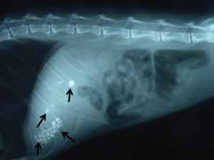
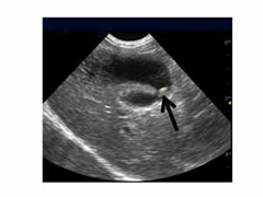
Radiographs (x-rays) are taken in animals with clinical signs and laboratory abnormalities consistent with biliary disease. Radiographs are useful in detecting stones in the biliary system and other abdominal disease that may be related to the biliary tract obstruction (Figure 1). The use of abdominal ultrasound is a very sensitive indicator of the cause the obstruction and should be performed in every animal suspicious of having an obstruction of the bile ducts or disease of the gallbladder (Figure 2).
Bile peritonitis is the inflammatory response of the lining of the abdominal cavity to the presence of free bile. Bile peritonitis is caused by rupture of the extrahepatic bile ducts, the gallbladder, or tears in liver lobes allowing bile to spill into the abdominal cavity. Rupture may be due to blunt trauma, neoplasia, a gallbladder mucocele, inflammation of the wall of the gallbladder, or obstruction from gallstones, cancer, or parasites. Your pet’s veterinarian may run tests comparing the bilirubin concentration of the abdominal fluid to the bilirubin concentration in the blood. Bile peritonitis is a surgical emergency.
The main goal of surgery is to confirm the underlying disease process, establish a patent biliary system, and minimize perioperative complications. Due to the complexity of biliary surgery, your veterinarian may refer you and your pet to an ACVS board-certified veterinary surgeon. Surgical options that your veterinary surgeon may discuss with you include:
- cholecystectomy – removal of the gallbladder
- cholecystotomy – incision into the gallbladder
- cholecystostomy tube – a tube placed into the gallbladder to provide drainage
- choledochotomy – an incision into the bile duct, usually to remove a stone
- choledochoduodenostomy – reattachment of the bile duct to a new location in the duodenum
- biliary-enteric anastomoses (cholecystoenterostomy) – attaching the gallbladder to the small intestine for permanent drainage
- choledochal stenting – placement of a temporary or permanent stent in the bile duct
- laparoscopic cholecystectomy – removal of the gallbladder using a laparoscopic technique
Patients having biliary surgery often require intensive care after surgery in a hospital with 24-hour nursing care available and may be hospitalized for days. Nutritional support is often needed with the placement of temporary feeding tubes. Pain medications and antibiotic therapy as well as liver targeted medications are often given.
Patients having biliary surgery have a high mortality rate (28–60%), though rates vary considerably depending on the causative condition.
Dogs with biliary tract obstructions are at an increased risk of acute kidney failure that develops due to the presence of bacterial endotoxemia. In dogs and cats, many authors have evaluated risk factors associated with outcome in patients undergoing surgery of the extrahepatic biliary tract. Factors besides kidney failure include:
- presence of septic bile peritonitis,
- elevated white blood cell count,
- prolonged clotting times,
- low blood pressure,
- sepsis, and
- disseminated intravascular coagulation.
The outcome for dogs and cats with bile peritonitis varies widely. If bacteria are present in the abdominal fluid (septic bile peritonitis) the prognosis is poor. If no bacteria are present in the abdominal fluid, the prognosis is favorable with surgical treatment, if the causative condition can be corected.
Surgical biliary intervention in cats with EHBO has about a 50% mortality rate overall and almost a 100% mortality when cancer is involved. Causes of death in these cats included clinical deterioration, bile leakage, and cardiopulmonary arrest. Cats who survive the initial surgery may experience long-term complications such as cholangiohepatitis (inflammation/infection of the liver), chronic weight loss, and recurrence of obstruction.
Premier Sponsors