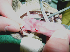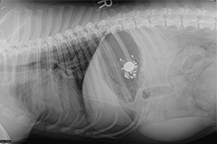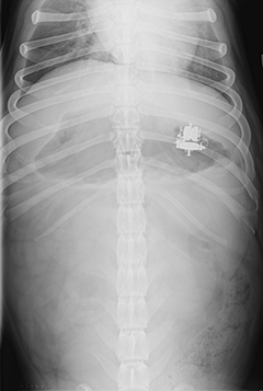

Gastrointestinal (GI) foreign bodies occur when pets consume items that are non-digestible and will not readily pass through their stomach or intestines. These items may be toys (Figure 2), leashes, clothing, sticks, or any other item that fails to pass, including human food products such as bones or trash. When these items become lodged in the GI tract, they can cause the animal to become very sick. The problems that are caused vary with:
- how long the foreign body has been present.
- the location of the foreign body.
- how badly the foreign material closes off the opening of the GI tract.
- what the material is made of. Some ingested items, such as older pennies or lead material, can cause systemic toxicities while others may cause regional damage to the intestinal tract itself due to compression or obstruction.
Gastrointestinal foreign bodies, especially strings, can sometimes cause holes in the tissue (or perforation). This causes spillage of intestinal contents into the abdomen. This condition quickly leads to life-threatening inflammation of the abdominal lining (peritonitis) and allows bacterial proliferation and contamination (sepsis). While some small foreign bodies will pass, many will become lodged along the gastrointestinal tract and cause discomfort and make your pet sick. Some foreign bodies located in the stomach may be retrieved with the use of an endoscope; however, most require surgical abdominal exploration and removal. Occasionally, foreign bodies will become lodged in the esophagus at the base of the heart or at the diaphragm, which may require thoracic (chest) surgery.


Clinical signs can vary significantly with the degree of obstruction, location, duration, and type of foreign body. Commonly noted signs include:
- vomiting
- anorexia (loss of appetite)
- abdominal pain
- dehydration
- diarrhea (with or without presence of blood)
In cases of linear foreign bodies, a string may be observed wrapped around the base of the tongue (Figure 1) or coming out of the anus.
The symptoms of a foreign body or intestinal obstruction can cause dehydration and electrolyte imbalances. This can cause the pet to feel systemically ill as well. Additionally, if the foreign body has perforated the GI tract and entered the chest or abdominal cavities, the animal may be profoundly ill and in critical condition. These may cause peritonitis, sepsis, and death. Many foreign bodies are made of materials that are potentially toxic when absorbed. Lead and zinc are good examples that when consumed may lead to profound systemic disease if enough is absorbed.

Your primary care veterinarian will likely recommend initial blood work that includes a complete blood count (CBC), serum chemistry, and a urinalysis. These combined will help to rule out other causes for your pet’s symptoms. Abdominal, and occasionally thoracic, radiographs are regularly performed (Figures 2 and 3). Positive contrast radiographs (using barium to highlight the inside of the stomach and intestines) may be performed when routine radiographs fail to show the cause for the clinical signs. Abdominal ultrasound can be very helpful in identifying gastrointestinal foreign bodies.
Surgery is not always required with gastrointestinal foreign bodies. Occasionally, the item ingested is small and smooth enough to pass through the gastrointestinal tract without causing damage or becoming lodged. Your veterinarian may recommend hospitalization for intravenous fluids and monitoring to help the item pass in this case. Additionally, some foreign bodies may become lodged in the upper gastrointestinal tract (mouth, esophagus, and stomach) and may be retrieved by making the pet vomit or with the use of a flexible endoscope (tube with a camera). If conservative management and endoscopy fail to provide relief, if the foreign body is not moving on x-rays, if the obstruction is getting worse, or if there is evidence of a linear foreign body, surgical exploration is warranted.
Esophageal foreign bodies require thoracic (chest) surgery to gain access for removal. Most gastrointestinal foreign bodies become lodged within the stomach or intestines and require a gastrotomy (opening the stomach) or enterotomy (opening the intestine). Once the item has been removed, the gastrointestinal tract is closed. Many linear foreign bodies and completely obstructed intestines are damaged severely enough that multiple enterotomies may need to be performed. If a section of bowel is irreversibly damaged, the section is removed and the healthy ends are reattached (an intestinal resection and anastomosis). The decision of which procedure to perform is determined by the surgeon when all the intestines and other abdominal organs have been evaluated.


