What is the Achilles’ tendon?
The Achilles’ tendon or common calcaneal tendon is made up of multiple tendons from several different muscles of the hind limb. The superficial digital flexor muscle and tendon, the gastrocnemius tendon and the combined tendon of the gracilus, semitendinosus and biceps femoris muscles are the main components.
What common injuries occur in the Achilles’ tendon?
A multitude of injuries can occur in the Achilles’ tendon, but there are two most common injuries:
Traumatic
- Breeds: Any
- Cause: lacerations, blunt force trauma, severe stretching/pulling
Atraumatic (chronic and degenerative in nature)
- Breeds: Any dog or cat but most common in Labrador Retrievers, Doberman Pinschers
- Cause: Repetitive injury, idiopathic (unknown), hypothyroidism, Cushings disease, Immune mediated polyarthritis
How can you tell if your animal has injured the Achilles’ tendon?
- Lameness or limping on the limb
- Crab claw stance (see Figures 1–3)
- Swelling around the injury (see Figure 4)
- Plantigrade (flat-footed or dropped stance) (see Figure 4)
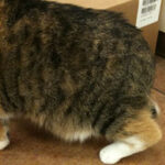
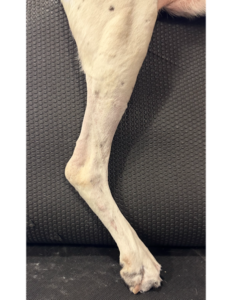
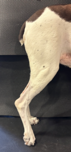
Figures 1–3: Claw-like stance in a cat and dog
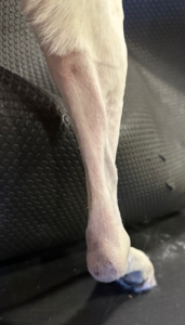
Figure 4: Dropped ankle stance and swollen tendon
Once the problem is localized to the Achilles tendon and ankle a series of tests may be performed to evaluate the tendon and the joint and determine then best course of treatment.
Physical exam
The physical exam is very important for diagnosing and localizing the injury to the tendon as well as identifying the level of discomfort.
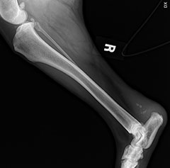
Radiographs (X-rays)
X-rays allow for evaluation of the ankle joint (tarsus) and the calcaneus (bone where the tendon attaches) to determine whether there is concurrent injury, the quality of the bone or arthritis associated with the degenerative disease. (See Figure 5)
Ultrasound
The ultrasound allows for direct visualization of the Achilles tendon in order to determine the extent and type of injury that is occurring. (See Figure 6)
CT
CT is considered a form of advanced imaging that allows for more detailed and 3-dimensional evaluation of the ankle joint and associated bones.
MRI
MRI is a form of advanced imaging that can be used to further evaluate the tendons, ligaments and soft tissues in and around the joint as well as the Achilles tendon.
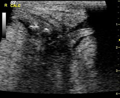
Figure 5: X-rays showing mineral in the tendon

Figure 6: Ultrasound showing injured areas in the tendon
Treatment options vary depending on your veterinarian’s or veterinary surgeon’s recommendations and the severity of the injury. Treatment options include:
External support (orthotics, casts, or splints)
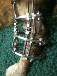
Surgery
This can involve attaching healthy ends of the tendon back together with suture, mesh, or other types of grafts. Most repairs to the tendon need to be protected after surgery to allow the tendon to heal. A variety of options exist for immobilizing the lower limb, including casts, splints, custom orthotics, temporary screws (Figure 4), linear external skeletal fixator, or a circular fixator (Figure 5).
Arthrodesis of the Tarsus (Ankle Fusion)
If there is complete disruption (tearing) of the Achilles tendon or severe damage to the ankle, joint fusion may be required for return to function. This entails stabilizing the joint in normal standing position using orthopedic implants (plates, screws, pins) and allowing the joint to fuse into one long bone. Joint fusion eliminates pain and lameness but does not restore normal flexion and extension of the joint.
Adjunct treatments
- Platelet Rich Plasma
- Extracorporeal shockwave therapy
- Stem cell therapies
Aftercare usually includes very restricted activity for 6–12 weeks post surgery and protecting the surgery repair with the aforementioned options for that time. Complete healing of the tendon can take a total of 9-12 months. If your veterinary surgeon has applied external support, regular appointments may be required to maintain and adjust the external support. Often times some element of physical therapy, either at home through the guidance of your surgeon or with a certified veterinary physical therapist, is indicated in order to re-establish an appropriate range of motion in the tarsus (ankle).
Potential complications include re-rupture or break down of the surgical site. These can often be avoided with appropriate postoperative care and restrictions.
Outcome
The prognosis is usually very good for the majority of injuries. Between 70-94% of dogs have a good to excellent return to function.
Premier Sponsors