The hyoid apparatus is a series of bones that suspend the larynx, or voicebox, and the tongue from the skull. The hyoid apparatus connects to the skull at the temporohyoid joint. This joint is where the paired, largest bones in the hyoid apparatus, the stylohyoid bones, articulate with the base of the skull. Temporohyoid osteoarthropathy, sometimes referred to as “THO”, or “middle ear disease”, is a disease associated with this articulation (the temporohyoid joint) from the hyoid apparatus and the skull. It is characterized by a proliferation of the bone surrounding the joint and which can lead to joint fusion.
The exact cause of THO is not completely understood, but may be due to either infection or inflammation spreading from nearby structures such as the inner ear or guttural pouch. Conversely, the disease may be a degenerative condition of the temporohyoid joint itself, somewhat similar to arthritis of other joints. The result of either infection and/or degeneration is bony proliferation and potential fusion the temporohyoid articulation. This decreases the normal range of motion and flexibility within these structures. Actions such as swallowing, movement of the head, neck or jaw area can cause a fracture within the attachment of the hyoid bones to the skull. The fracture can cause rapid worsening of clinical signs as well as compression of the facial and vestibulocochlear nerves which are responsible for facial expression, eye lubrication, and balance (cranial nerves 7 and 8).
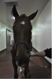
Signs can be vague and vary from horse to horse. Some early signs include pain when pressure is applied to the base of the ear or at the throatlatch area, tossing the head or reluctance to accept the bit or perform under saddle in specific head positions. Once there is significant thickening of the bones, or a fracture has occurred, more marked signs including loss of coordination (ataxia), lip and ear droop (Figure 1), tilting of the head to one side, or difficulty swallowing can occur. Most of these signs are due to nerve damage in the affected area (cranial nerves 7 and 8). Damage to the nerves supplying function to the eye can result in decreased tear production or an inability to blink appropriately; this can lead to serious corneal ulceration.
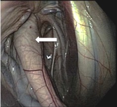
There are several different ways that THO can be diagnosed. Your primary care veterinarian can help guide you in choosing the best diagnostic plan for your horse. Skull radiographs, or x-rays, can detect the characteristic thickening of the affected bones but often are not definitive. Upper airway endoscopy, or “scoping” can often confirm the diagnosis. Your veterinarian can use the endoscope to see inside the guttural pouch, where the stylohyoid bone can be observed as it articulates with the skull. Horses with THO have an enlargement of the stylohyoid bone in this area, as seen in Figure 2. It is important to check both guttural pouches because the disease can be bilateral (affecting both right and left sides).
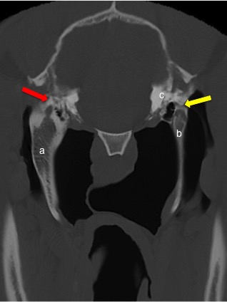
Computed tomography, or CT scans, can provide significantly more detailed information about the disease, detecting both bony and soft tissue changes within the hyoid apparatus, skull, and inner ear (Figure 3). This level of detail is often helpful for guiding any surgical procedures (see below).
Medical therapy includes antibiotics to treat any infection of the inner ear and anti-inflammatory drugs to decrease both pain and inflammation. It is important to diagnose and treat corneal ulceration that may have occurred as a result of any decreased tear production or blinking. Often treating the corneal ulcers requires long term topical antibiotic treatment, potentially combined with a tarsorrhaphy (temporary surgical closure of the eyelids to protect the cornea) or other medical therapy as recommended by your veterinarian.
A ceratohyoidectomy is a surgical procedure that has been developed to treat THO. This involves removing one of the small bones of the hyoid apparatus, the ceratohyoid bone, completely. It is designed the decrease the force applied from the hyoid apparatus to the skull and decrease the risk of fracture; this also decreases the pain and discomfort associated with the abnormal or fused joint. Unlike previous surgical treatments, regrowth or recurrence of clinical signs has not been noted as a complication. An ACVS board certified veterinary surgeon can help determine if this procedure is appropriate for your horse.
The prognosis for improving clinical signs is generally considered favorable, with most horses able to return to their original use. However, existing nerve damage present prior to surgery may persist, and some horses have small nerve deficits (such as a head tilt or lip and ear droop) that may remain after treatment. If diagnostics indicate that a fracture has already occurred, the prognosis may be lower depending on the degree of neurologic signs present.
The aftercare for your horse will vary depending on the severity and nature of the signs present. The duration of neurologic recovery will vary and usually takes several months, however, in some cases improvement may take over a year. If corneal ulceration is present due to decreased tear production, or decreased blinking, this may also require extended treatment though complete resolution occurs in the majority of cases.
Premier Sponsors