A very common source of sudden onset lameness in horses is the hoof abscess. Almost every horse owner will experience this situation at one time or another; their horse is standing, pointing or resting a leg, unwilling to place much weight on it. Sometimes there is so much pain, the horse is unwilling to stand on the leg at all and often times this occurs suddenly (i.e. they were ‘fine’ earlier today or yesterday). The horse may be trembling or sweating, as some of these abscesses can be very painful.
The source of pain is infection, trapped between the confines of the hard hoof wall and hard coffin bone. The pressure from the infection and swelling of the soft tissues/laminae makes this condition very painful. The majority of these conditions are very easily handled with prompt medical attention by your primary care veterinarian, although in a small number of cases, it may require more aggressive surgical intervention.
Your horse may exhibit one or more of the following signs:
- Sudden onset lameness
- Elevated/increased digital pulse
- Warm/hot hoof
- Fever (>101.5°F)
- Cellulitis/swelling of the lower limb, above the affected hoof
- Standing, pointing, or resting a leg
- Unwilling to place weight on a leg
- Trembling or sweating with an elevated heart rate
- Positive response to hoof testers
Your veterinarian may:
- Perform a physical examination
- Clean out the hoof
- Use hoof testers around the hoof to reveal a localized area of intense sensitivity to pressure
- Remove a shoe, if one is in place
- A hoof knife may be used to carefully open the abscess through the sole/bottom of the foot, providing drainage of pus and allowing for the area to be soaked, medicated, and bandaged.

As with most medical conditions, there are instances where the situation may become more complicated. Sometimes, the infection in the foot doesn’t form a discrete abscess or if the abscess is not resolved in a timely manner, it may course within the hoof and infect the coffin bone directly (osteomyelitis). In these situations, simply draining the abscess and treating with antibiotics will not cure the infection because it has invaded an area of the coffin bone.
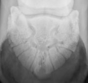
Progression from a straight forward hoof abscess to osteomyelitis can be suspected when the abscess and lameness don’t respond in the usual time frame or there is continued drainage for a prolonged amount of time. Radiographs can be used to confirm the diagnosis (Figure 1). Surgery is then required to remove the infected bone and associated soft tissues.
Medical:
- The area of sensitivity and underlying abscess is carefully opened by your veterinarian to allow for drainage, soaking, direct application of medication, and bandaging.
- Antibiotics and anti-inflammatory medication may also be prescribed.
- Once the abscess stops draining and resolves, the area should heal and cornify- meaning the healing tissue produces horn to become tough like the rest of the foot. The shoe can be replaced once this has occurred and the horse can go back out without a bandage and go back to work.
Surgical:
- An ACVS board-certified veterinary surgeon is experienced and skilled with handling osteomyelitis of the coffin bone and will have specialized tools and methods that effectively and quickly resolve the infection minimizing downtime and discomfort to the horse.
- Surgical debridement of the coffin bone is generally handled as a standing surgery in a hospital setting, with the horse sedated, under regional anesthesia (local blocks) of the foot and with the aid of a tourniquet.
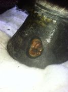
- A motorized burr (Dremel tool) is used to bore a small hole in the hoof wall over the lesion (hoof wall resection). Figure 2.
- Approaching the bone infection through the hoof wall rather than through the sole allows complete access to the area and provides for immediate post-surgical comfort. Approaches through the sole can result in tremendous post-procedure pain and the requirement for special shoes with treatment plates and bandages.
- The horse may be treated with systemic antibiotics and antibiotics delivered by regional limb perfusion (RLP). The antibiotic treatment is guided by bacterial culture of the infected tissue taken at surgery. With an RLP, a tourniquet is used to isolate the digit and a high dose of antibiotics is delivered into a vein or artery in that isolated area. Very high tissue levels of antibiotics can be achieved in this way. Because peak tissue levels of antibiotics determine their effectiveness and because RLPs allow much higher antibiotic levels in the local tissues compared to other methods (oral, intravenous) RLPs have proven to be a very good way to treat localized bone infections. The procedure is usually performed several times in the days following surgery.
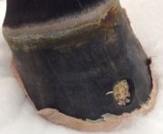
- Another great tool that may be used after surgical debridement of the coffin bone is the use of fly larvae that are produced specifically for this purpose. Medical Maggots™ can be placed into the surgical site to complete the removal of diseased tissue after surgery has been performed. They are sterilely produced for the purpose of debridement of necrotic tissue in wounds. The fly larvae only consume dead and necrotic tissue and do so at a very minute level (Figure 3).


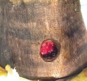
After hoof wall resection and surgical debridement of the infected coffin bone, the foot is kept under a sterile bandage, changed every 48 hours until the wound bed cornifies (Figure 4). Antibiotics and anti-inflammatory medications may be continued based on culture and sensitivity results in addition to the progress of the horse. After this, a lighter bandage is maintained simply to keep debris out of the defect. The hoof wall defect will grow out over a couple of months. In general, the hoof wall grows at a rate of about one centimeter each month. Once the hoof wall grows out, the foot is generally as good as new.


