The thyroid glands are paired structures located along the windpipe (trachea), about halfway down the neck of dogs and cats. The thyroid glands are responsible for producing hormones that are vital for normal body function. Thyroid growths in dogs can be benign (adenoma) or malignant (carcinoma). Benign growths tend to get larger and may produce excess hormones; malignant growths can also spread to other parts of the body. While benign tumors of the thyroid gland are common in cats, the majority of dogs have malignant tumors. Thyroid tumors are commonly seen in middle aged to older large breed dogs such as boxers, beagles, golden retrievers, and Siberian huskies.
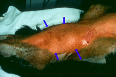
Dogs with thyroid tumors may have no symptoms or may develop a lump in the neck region (Figure 1). If the mass compresses the windpipe (trachea), these dogs may present with difficulties breathing or coughing. If the mass is pressing on the esophagus your dog may gag or have difficulty swallowing. Some dogs may have a change in their bark, weight loss, or loss of appetite. Although the majority of canine tumors are malignant, they rarely produce excessive hormones that are associated with clinical signs of hyperthyroidism, such as:
- restlessness
- increased appetite
- weight loss
- hair coat abnormalities
- drinking and urinating more than usual
Thyroid tumors can occasionally be found in a location in the neck away from the normal thyroid glands or even under the tongue or in the chest.
Thyroid masses occasionally cause neck swelling which can be seen on radiographs (x-rays) but other imaging techniques such as ultrasound or computed tomography (CT) scan are better for assessing the size and invasiveness of the tumor. Definitive diagnosis is based on microscopic examination of a tissue sample. Due to the highly vascular nature of the tumor, coagulation parameters should be assessed using blood clotting tests prior to biopsy or surgery.
Additional testing is usually performed before surgery to stage the tumors. Chest x-rays or CT, abdominal ultrasound, and blood work are used to investigate whether there is evidence of thyroid hormone production or metastasis (spread of the cancer), and assess other organ function.
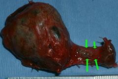
Removal of these tumors can be difficult because the tumors can invade local blood vessels or other tissues (Figure 2). Because tumors that are large or invasive can be difficult to remove, referral to an ACVS board-certified veterinary surgeon is indicated for any large or fixed tumors. Radiation or chemotherapy is often recommended for masses that are incompletely resected or are too large for surgical removal.
Radioactive iodine (I-131) treatment has been shown to be an effective adjunct to the treatment on thyroid tumors. I-131 can be utilized in patients that are poor surgical candidates or in patients where vascular invasion has been identified in spite of surgical removal.
Pathologist review of the removed tumor is important to determine if these additional treatments (chemotherapy, radiation therapy or I-131 treatment) would be beneficial for your pet Pathologic evaluation of the removed tissues may include special stains (immunohistochemistry) that helps veterinarians tailor follow up treatment of the tumor.
After surgery your dog may have a soft bandage around his neck. You should avoid putting any leashes or collars around your dog’s neck until they have healed from surgery, usually 10–14 days. Use a body harness for walking your pet instead. During this time period you should keep your dog’s activity limited.
Follow the advice of your veterinary surgeon regarding medications that may be needed for your pet after surgery. If both thyroid glands are removed, your veterinarian may need to check your dog’s calcium levels several times during recovery because some parathyroid tissue is removed with the thyroid glands (parathyroid glands are involved in calcium regulation).
Surgical removal of thyroid tumors has the best outcome if the mass is freely moveable, less than 4cm in size, nonmetastatic (has not spread) and can be completely removed. Long-term survival (1 to 3 years) may be achieved in dogs, depending on histologic features observed by the pathologist and early diagnosis prior to local invasion or metastatic (spreading) disease. Patients treated with surgery and follow-up I-131 treatment have an average survival of 34 months.
There are always risks associated with general anesthesia. Complications specific to removal of thyroid tumors in dogs include bleeding or damage to the recurrent laryngeal nerve, which is responsible for movement of the larynx (upper airway cartilages) during breathing and swallowing. Dogs that have both thyroid glands removed may experience low calcium (hypocalcemia) or low thyroid hormone levels (hypothyroidism) which require treatment with medication. Finally, there can be some swelling of the incision on the neck after surgery.
Premier Sponsors


