
Splenic masses can be cancerous or non-cancerous processes. Splenic hematoma and nodular hyperplasia are the most common non-cancerous lesions found in the spleen and account for 20–41% of all splenic lesions. They are benign nodules/masses of clotted blood. Surgical removal is curative.
Cancerous masses can cause internal bleeding that is not explained by trauma. The majority of cancerous masses—up to two-thirds of masses on the spleen—are called hemangiosarcoma. Hemangiosarcoma is usually seen in older dogs, typically 8–10 years of age. Large-breed dogs appear to be at an increased risk, especially German Shepherds, Golden Retrievers, Labradors, and Poodles.
Signs associated with splenic masses can be subtle and include weakness or may be more obvious e.g. collapse and sudden death if the mass ruptures and bleeds internally. Pets may have pale gums, increased heart rate, or increased respiratory rate. Other signs can include
- Swelling of the abdomen
- Weight loss
- Loss of appetite
- Fainting or weakness
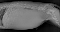
Figure 1. Radiograph of a dog showing a large splenic mass occupying most of the abdomen.Your primary care veterinarian may run several tests to obtain a presumptive diagnosis and to prepare for surgery. These may include blood tests, urinalysis, a clotting profile, examination of fluid obtained from the abdomen, chest, and abdominal radiographs (Figure 1). Abdominal ultrasound is another useful method to identify and characterize masses in the abdomen as well as look for free fluid or blood. Echocardiography (ultrasound of the heart) may be recommended as some dogs may have tumors spread to the heart. The only way to definitively determine if a splenic mass is non-cancerous or cancerous, is by removal of the spleen and analysis of the tissues under a microscope. Needle aspirate often is unable to determine these.
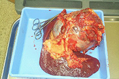
Surgery is the primary method of treatment for dogs with splenic masses. This involves the removal of the spleen (splenectomy). Removal of the spleen is preferred to a biopsy as it serves as both a diagnostic and therapeutic procedure (Figure 2). If the splenic mass has ruptured and is causing internal bleeding, this procedure must be done as soon as possible to prevent further blood loss. Patients are stabilized prior to surgery. This may require fluid therapy or a blood transfusion and intensive care monitoring.
Most animals will remain in the hospital for 24–48 hours after surgery to monitor for heartbeat changes and assess their blood volume. Your pet will have a large incision on their abdomen, and after surgery, activity should be restricted to short leash walks only during the first two weeks of healing. Your dog may need to wear an E-collar or tee shirt to prevent self-trauma to the surgical site.
Prognosis depends on the outcome of the biopsy. Benign splenic masses are effectively cured with surgery. Unfortunately, up to two-thirds of dogs with splenic masses have a cancerous mass and most of these masses are an aggressive tumor (hemangiosarcoma). This tumor rapidly spreads to other areas of the body and is not responsive to chemotherapy. Survival times with surgery alone for dogs with hemangiosarcoma may be two–three months or less. One year survival is less than 10%. Ultimately, dogs die from metastatic disease. Chemotherapy may increase survival times up to six–eight months.
Complications that may be associated with surgery include hemorrhage (ongoing bleeding), cardiac arrhythmias (irregular heart rhythm), and pancreatitis (often manifested by vomiting). An ECG to look for arrhythmias is recommended after surgery. While this may require treatment, most arrhythmias are resolved within 24–48 hours.
Photos provided courtesy of Elizabeth Hardie, DVM, PhD, Diplomate ACVS.
Premier Sponsors