The most common type of primary liver tumor, hepatocellular carcinoma, originates from liver cells (hepatocytes) and has a low rate of spread to other organs. Other types of primary liver tumors can originate from:
- bile ducts
- connective tissue
- blood vessels
- hormone-secreting cells (neuroendocrine)
Metastatic liver tumors are those that have spread from another organ of the body to the liver. Tumors that spread to the liver from another organ or region of the body are more common than those that originate within the liver itself.
When a large, single mass is located in the liver, called a massive tumor, a hepatocellular carcinoma is the diagnosis in at least half of dogs. Cats tend to develop more benign tumors than dogs. Bile duct adenomas (biliary cystadenomas) account for more than half of all liver tumors in cats, yet are uncommon in dogs. Bile duct carcinomas are the most common malignant liver tumor in cats and the second most common liver tumor in dogs. Uncommon liver tumors include:
- carcinoids
- sarcomas of various types
- myelolipomas
Signs of a liver tumor are typically nonspecific and usually do not point to the liver as the primary source of the problem. Approximately 75% of dogs and half of cats show signs of illness, including:
- decreased appetite
- weight loss
- lethargy
- vomiting
- increased thirst
- increased urination
- distention of the abdomen with fluid
- seizures
Some dogs and cats become jaundiced, which is a visible yellow discoloration of the whites of the eyes, gums, inner ear flaps and poorly haired areas of skin.
Your primary care veterinarian may detect a liver mass upon palpation of the abdomen. Further testing may include:
- a blood analysis (complete blood count, chemistry profile, coagulation profile and urinalysis)
- abdominal x-rays
- abdominal ultrasound
- chest x-rays
- advanced imaging such as computed tomography (CT) scan or magnetic resonance imaging (MRI)
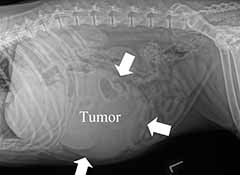
Based on abdominal x-rays or abdominal ultrasound, your pet’s veterinarian may make a presumptive diagnosis of a liver tumor. (Figure 1) Due to the complexity of liver surgery and the risk of bleeding complications during surgery, many primary care veterinarians will refer your pet to an ACVS board-certified veterinary surgeon for removal and post-operative care.
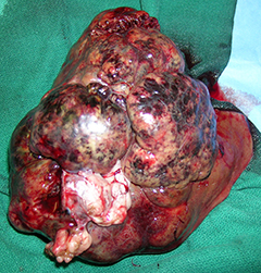
The treatment of choice for primary liver tumors is surgical removal. A large incision is made into the abdomen, and the affected liver is removed with surgical ties or a stapling device. For very large tumors, a second incision along the rib cage may be required to have enough room to get to the tumor. Sampling of any other abnormal findings in the abdomen is also performed as needed.
Bleeding is a risk of liver surgery. Your surgeon will likely ensure that there are blood products available for a blood transfusion for your pet if there is a large amount of bleeding in surgery. Another complication of surgery is failure to remove all abnormal tissues or leaving “dirty margins”. This can be a function of where the tumor is growing on the liver or the lobe that is affected.
After surgery, your pet is monitored in an intensive care unit and medication is administered to control pain. Fluids and antibiotics may be administered via an intravenous catheter for a few days after the procedure to maintain hydration and prevent infection. Daily blood tests may be performed to check for signs of internal organ dysfunction and internal bleeding. If needed, a blood or plasma transfusion may be given.
At home, pain medications and antibiotics may be required. If your pet does not eat a regular diet, a low fat home-cooked diet may be offered. An Elizabethan collar (i.e., a protective device worn around an animal’s neck) is kept on your pet when not under your direct supervision to prevent licking of the incision. Alternatively, a tee shirt can be put on your pet to protect the incision. Pet owners should schedule a recheck evaluation with their pet’s veterinary surgeon about 10–14 days after surgery.
Long-term prognosis after liver tumor removal depends on the tumor type, completeness of removal, and presence of spread (metastasis). The outcome of surgical treatment of hepatocellular carcinomas is generally very favorable, with survival times commonly exceeding 3.8 years and metastasis seen in less than 5% of pets. Removal of resectable biliary cystadenomas in cats has a good prognosis with long survival times. Surgical removal of bile duct carcinomas yields short survival times in both dogs and cats due to metastasis and regrowth of the tumor in the liver. Sarcomas and carcinoids have a poor prognosis, as the majority of these have metastasized at the time of diagnosis.
Dogs that have untreated primary liver tumors (specifically hepatocellular carcinoma) are 15 times more likely to die of tumor-related complications than dogs that have had their tumors removed. Liver tumors are fragile and may rupture at any time, thus may cause life-threatening internal bleeding. A tumor may compress the main bile duct that drains bile from the liver into the intestine, thereby causing jaundice. It may compress internal organs or the great vessels in the abdomen and cause a variety of signs, such as vomiting and distention of the abdomen with fluid. Rarely, liver tumors produce insulin-like substances that cause the pet to have low blood sugar.
Ongoing bleeding after surgery, although seen in less than 2% of operated patients, can result in death of the pet in the postoperative period. Other complications may include:
- infection
- twisting of a liver lobe adjacent to the portion of liver that has been removed
- re-growth of the tumor in the liver
- spread of the tumor to other internal organs
Premier Sponsors


